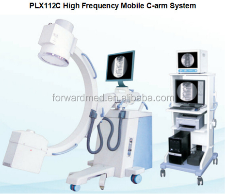
fluoroscopy C-arm x ray machine prices /x-ray x-ray prices photographic film x-ray price PLX112C

1 Set
MOQ
fluoroscopy C-arm x ray machine prices /x-ray x-ray prices photographic film x-ray price PLX112C
Features Gallery Product Description Request A Quote
Features
Specifications
Model Number: PLX112C
Properties: Medical X-ray Equipments & Accessories
Output Power: 5kw
Frequency: 40kHz
Tube Current: 40 - 120 KV
Continuous Fluoroscopy Mode: 0.3mA~~4mA
Spotfilm: N/A
Radiography: 20-100mA
Pulse Fluoroscopy: 4.1mA~~30mA
Image Intensifier: Toshiba
Filed Viewing: 9"
LCD Display: 2 Units 19" Medical LCD Display, 1 Unit 14" CRT Monitor
Basic Infomation
Model Number: PLX112C
Payment & Shipping Terms
Packaging Details: Standar Export Wooden Case
Supply Ability: 200 Set/Sets per Month
Product Description

fluoroscopy C-arm X Ray/ X-ray Price C-arm X Ray /x-ray machine prices photographic film X-ray Price

Specifications:

Fluoroscopic Capacity Max rated capacity :

Tube Current 4mA, Tube Voltage 120kV

Automatic Fluoroscopy:

Tube Voltage: 40kV~120kV adjust automatically,Tube Current : 0.3mA~4mA set manually

Manual Fluoroscopy :

Continuous Tube Voltage:40kV~120kV, Continuous Tube Current : 0.3mA~4mA

Pulse Fluoroscopy :

Continuous Tube Voltage 40kV~120kV , Continuous Tube Current 4.1mA~30mA

Pulse frequency: 0.1~25fps,continuously,adjustable

Photography Capacity: Max rated capacity : 5 KW

Tube Voltage and Current Combination :

40kV~49kV 1 ~180 mAs

50kV~59kV 1 ~140 mAs

60kV~69kV 1 ~125 mAs

70kV~79kV 1 ~110 mAs

80kV~89kV 1 ~100 mAs

90kV~99kV 1 ~80 mAs

100kV~109kV 1 ~63 mAs

110kV~120kV 1 ~50 mAs

Plateholder Size: 200mm×250mm(8″×10″) or 50mm×300mm(10″×12″)

X-ray Tube Special for High Frequency

Fixed anode Dual-focus : small focus: 0.3mm, large focus: 1.5mm

Inverter Frequency: 40KHz

Thermal capacity : 35KJ (47HU)

Video System :

mage Intensifier : TOSHIBA 9″ Image Intensifier

CCD Video camera : One million pixels ultra low-light CCD camera imported from Japan.

Monitor : High-definition, high-resolution, optional specification

Image Processing System: (standard) : real-time image collection, specular image , recursive level 0-15 continuously adjustable, last frame frozen, Gamma correction, mass storage, image W/L adjustable, gray-scale conversion,interest district balance, reversal, noise reduction,intensify, smooth, sharpen, compress, enlarge, measurement, label, print and typesetting, DicomSCP, DicomSCU, DicomDIR, Dicom Printing

Structure Directive wheel : ±90°revolution, can freely change the moving direction of the unit.

Ascending & Descend- ing Range of Pillar : ≥400mm

C-arm : Forward and Backward Movement: 200mm,

Revolution around Horizontal Axis: ±180°

Revolution around Vertical Axis : ±15°,

Focus screen distance:1000mm,

C-arm opening :800mm,

C-arm arc depth : 640mm

Slip on orbit: 120°(+90°~ -30°)

Packing Details: 2400mm×1100mm×1420mm N.W. 300kgs G.W.400kgs

Usage:

Orthopaedics: osteopathy, diaplasis, nailing

Surgery: removing foreign body, cardiac catheter, implanting pace maker, interventional therapy, partial radiography, local photography, and other work.

Features:

1. Leading domestic level Mobile Surgical X-ray machine. Real continuously pulse fluoroscopy 25fps. Convenient connection with the Digital subtraction system.

2. One million pixels CCD camera makes the image more distinct.

3. World-class image processing technology software makes the image more distinct to make it more convenient for the doctors in the operation and diagnostic process. Standard DICOM interface makes it convenient to connect with the hospital information system.

4. The pulse fluoroscopy can obtain high –definition image from low dose to meet the requirement of the high-precision and high difficulty microinvasive operation

5. Low radiation dose used can protect the doctor and patient.

6. Unique double foot brake controller design makes it convenient to control the instrument inside and outside the operation room. This design can also protect the health care professionals by reducing the opportunity to come into contact with the radiation.

7. Exclusive inside and outside monitor design makes it more convenient to observe the operation process either inside or outside the operation room. At the same time, this design can facilitate the outdoor teaching and the real-time observation of the surgical procedures.

8. Uniquely designed basement electric auxiliary supporting arm, ensuring safer use.

9. Specially designed portable controller, facilitating operation.

10.New-pattern frame design with a small and pleasing shape.

11.High-quality combined HF-HV X-ray generator, greatly reducing the X-ray irradiation.

12.The fluoroscopic KV and MA automatic tracking function makes the image brightness and definition shift to the best condition automatically.

13.Toshiba image intensifier is employed, stable and reliable in quality and sound in image definition.

14.Dense X-ray grid is used, further improving the image definition.

Recommended Products
Get in touch with us
Contact Person : Allen
Characters Remaining(20/3000)